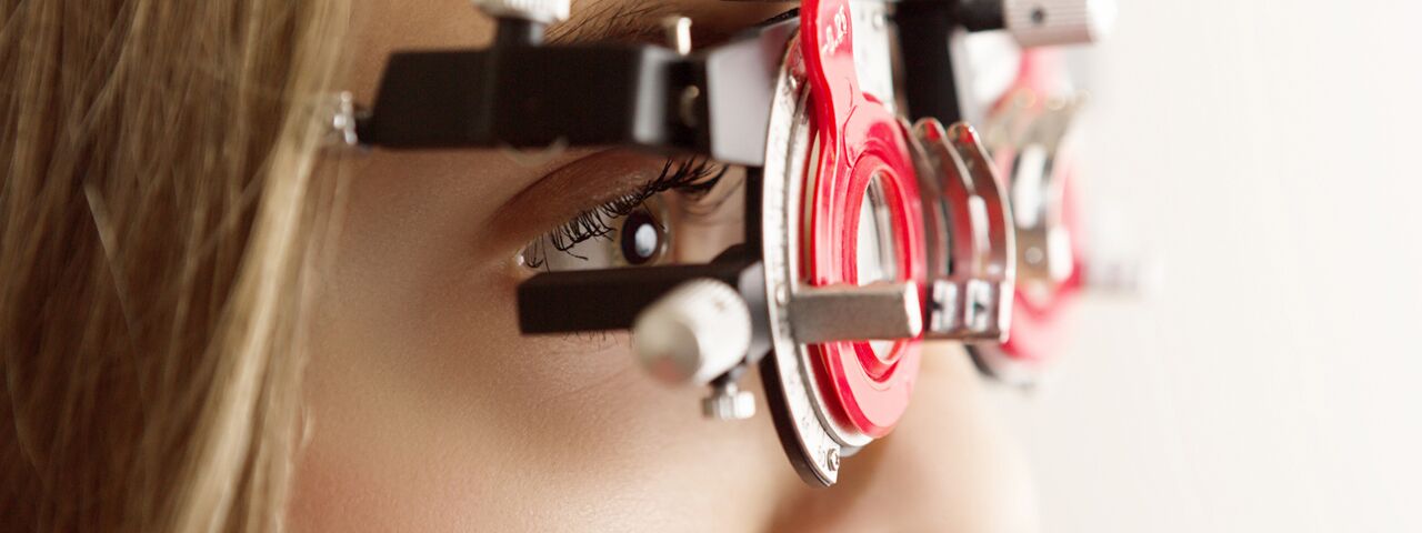
We use the most up-to-date technology to ensure the best eye care possible. Here are some of the different types of tests and equipment you may experience on a visit to our Practice.
RETeval®
 The RETeval® device is opening up access to functional information through visual electrophysiology testing in a whole new way. The device is the first completely portable, handheld, full field flash ERG and VEP testing device. We can now get the full picture of patient eye health with non-invasive, effective and reliable testing – leading to better patient care and outcomes.
Perfect for pediatric patients and adults alike, the RETeval device offers valuable information for a more effective diagnosis and monitoring of vision-threatening diseases.
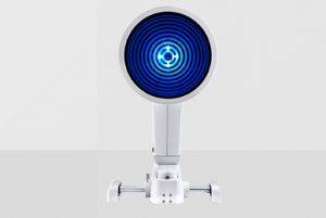
RAPDx
 RAPDx utilizes a high-definition, machine-vision system under controlled infrared conditions to present monocular visual stimuli while recording binocular pupil responses. Unlike human observers that only see one eye response at a time, RAPDx simultaneous images both direct and consensual light responses. The power of the system is to collect a series of responses and average them into a consolidated response curve. Pupil testing is notoriously non-standardized, variable, and noisy. Response averaging significantly helps filter the noise inherent in the biological responses.
Easy, objective, quantitative, delegated, detailed … perhaps better than the finest human observer. RAPDx may be the most significant advancement in pupil testing in decades.
Sensory Cradle
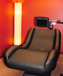 Designed to help adults and children meet their visual cognitive, academic and developmental goals, the Sensory Cradle Program incorporates Vision, Proprioception and Auditory Sensory Modalities to help re-train the brain. A 30 day process that consists of three phases, the sensorimotor integration technique is designed to help individuals with the following conditions:
- Autism, ADD/ADHD, Trauma/PTSD
- Sensory Integration Disorder, Traumatic Brain Injury, Balance/Coordination
- Acquired Brain Injury, Developmental Delays, Vision/Concentration
- Behavioral Problems, Anxiety/Depression
Read more about the Sensory Cradle
ColorDx Test
 Up to 15% of the general public may have a deficiency in the way they see the world’s colors. While colorblindness is genetic, color deficiencies can be caused by many kinds of retinal, macular, optic nerve, trauma or neurological disorders. Most tests can only detect genetic color vision deficiencies and late stages of any one of the other disorders.
The ColorDx test allows for a more nuanced and numerical approach to color deficiency. Giving you the confidence that you are seeing the world the way you want to see it.
Corneal Mapping
Corneal topography, also known as photokeratoscopy or videokeratography, is a non-invasive medical imaging technique for mapping the surface curvature of the cornea, the outer structure of the eye. Since the cornea is normally responsible for some 70% of the eye’s refractive power, its topography is of critical importance in determining the quality of vision.
 Keratograph
 Keratograph
The OCULUS Keratograph® 5M is an advanced corneal topographer with a built-in real keratometer and a color camera optimized for external imaging. Unique features include examining the meibomian glands, non-invasive tear film break-up time and the tear meniscus height measurement and evaluating the lipid layer.
Digital Retinal Imaging & OCT Scans
Digital Retinal Imaging
Digital Retinal Imaging allows your eye doctor to evaluate the health of the back of your eye, the retina. It is critical to confirm the health of the retina, optic nerve and other retinal structures. The digital camera snaps a high-resolution digital picture of your retina. This picture clearly shows the health of your eyes and is used as a baseline to track any changes in your eyes in future eye examinations.
Optical Coherence Tomography (OCT)
 An Optical Coherence Tomography scan (commonly referred to as an OCT scan) is the latest advancement in imaging technology. Similar to ultrasound, this diagnostic technique employs light rather than sound waves to achieve higher resolution pictures of the structural layers of the back of the eye.
A scanning laser used to analyze the layers of the retina and optic nerve for any signs of eye disease, similar to an CT scan of the eye. It works using light without radiation, and is essential for early diagnosis of glaucoma, macular degeneration and diabetic retinal disease.
An OCT scan is a noninvasive, painless test. It is performed in about 10 minutes right in our office. Feel free to contact our office to inquire about an OCT at your next appointment.
Visual Field Testing
A visual field test measures the range of your peripheral or “side” vision to assess whether you have any blind spots (scotomas), peripheral vision loss or visual field abnormalities. It is a straightforward and painless test that does not involve eye drops but does involve the patient’s ability to understand and follow instructions.
An initial visual field screening can be carried out by the optometrist by asking you to keep your gaze fixed on a central object, covering one eye and having you describe what you see at the periphery of your field of view. For a more comprehensive assessment, special equipment might be used to test your visual field. In one such test, you place your chin on a chin rest and look ahead. Lights are flashed on, and you have to press a button whenever you see the light. The lights are bright or dim at different stages of the test. Some of the flashes are purely to check you are concentrating. Each eye is tested separately and the entire test takes 15-45 minutes. These machines can create a computerized map out your visual field to identify if and where you have any deficiencies.
OPTOS Retinal Exam
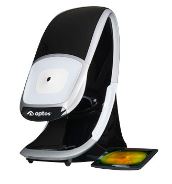 Annual eye exams are vital to maintaining your vision and overall health. We offer the optomap® Retinal Exam as an important part of our eye exams. The optomap® Retinal Exam produces an image that is as unique as you fingerprint and provides us with a wide view to look at the health of your retina. The retina is the part of your eye that captures the image of what you are looking at, similar to film in a camera.
An optomap® Retinal Exam provides:
- A scan to show a healthy eye or detect disease.
- A view of the retina, giving your doctor a more detailed view than he/she can get by other means.
- The opportunity for you to view and discuss the optomap® image of your eye with your doctor at the time of your exam.
- A permanent record for your file, which allows us to view your images each year to look for changes.
Please schedule your optomap® Retinal Exam today!
For more information on the optomap® Retinal Exam, go to the Optos website .
*Saturday hours bi-monthly, please check with office to confirm we are open.